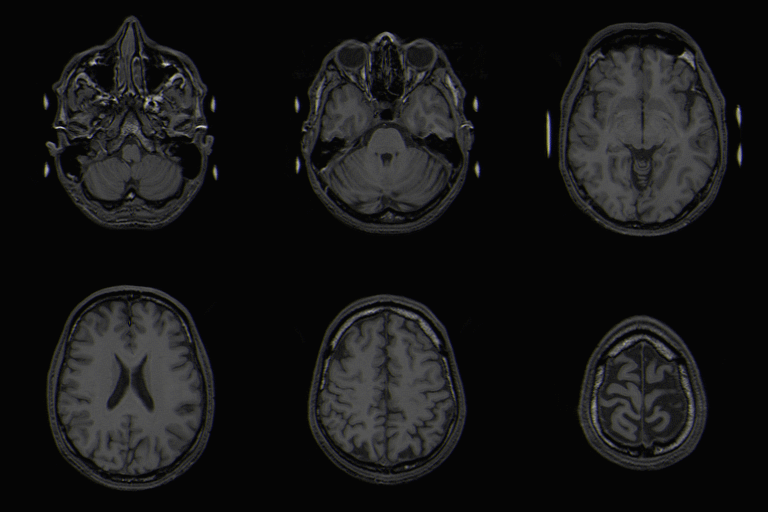
Category:Neuroimaging
Contents
Neuroimaging
There are have been many differences observed between typically developing children and those with ASD in neuroimaging studies using fMRI, EEG, transcranial magnetic stimulation, EMG, and structural MRI. It has been hypothesized in the Mirror Neuron System Theory of Autism that the social/communication deficits in ASD arise because of differences in activation of the Mirror Neuron System (MNS), since the mirror neuron system plays an integral role in mediating understanding of emotional states of others. However, many different areas of the brain can participate in complex social/communicative tasks, making it difficult to single out just one neural area responsible for the widespread abnormalities in socialization and communication experienced by patients with ASD. Much of current neuroimaging research in ASD operates under one of two different camps that attribute autism to different areas of less activation in the brain.
- Theory of Mind-Patients with ASD show lower or no activation in midline structures as compared to a control groups in many tasks, lending credence to the 'Theory Of Mind' explanation for social/communication deficits in patients with ASD. In this theory, patients with ASD have lower activation of mirror neurons in the midline structures which are responsible for "reflecting" about others' emotions or wants1,. Thus, patients have social/communication deficits because they have are unable to process the meaning of other peoples' emotions, which suggests a problem in executive processing.
- Embodied Simulation Theory- Scientists who want to test this theory focus their research on shared circuits that are involved in both one's own emotions and other's emotions. Numerous studies have shown that when subjects witness a goal oriented task many different types of neurons are activated in addition to those in the midline structures. For example, when a person watches another person drink a glass of milk with an expression of disgust on his face, the premotor and parietal areas are activated for the action itself, the insula areas are activated in response to the emotion of disgust, and the secondary somatosensory area is activated for the sensations involved in the task. These are the same areas that would have been activated if we drank the milk and were ourselves disgusted. Thus, we are able to understand others' emotional states and intentions because contextual cues activate the same neural circuits that are activated when we perform the same task. However, patients with ASD show diminished activation of these areas as compared to control groups. So, in this theory, the social and communication difficulties arise in patients with ASD because these areas are less activated. Consequently, they have difficulty "understanding" another's actions.1
These two theories are not entirely incompatible and there have been some hypothesis on how the two theories could be integrated to provide a better explanation for the differences seen.
Neurosystems affected in ASD
Neuroimaging has identified several neural systems that map onto various components of the sympomatology seen in Autism Spectrum Disorders. Arousal, reward, and face processing systems demonstrate the potential utility of neural systems knowledge.
The arousal and reward systems are likely candidates that help a person determine the social significance of stimuli, which is an impairment seen in those with autism. These two systems are likely to be involved in regulating social communication. The facial processing system has also been shown to be abnormal in individuals with ASD, possibly due to reduced experience and subsequently ability in perceptual processing of faces. An eye-tracking study on the social phenotype in autism found that facial processing difficulties were tracked back to difficulties in determining what is socially salient in naturalistic social scenes, which may be caused by abnormal arousal and reward responses to faces in those with ASD. 4
Several fMRI studies have indicated that the amygdala is a signficant contributor to social processing in typically developing participants and to the social processing impairment in autism because of the amygdala's role in arousal processes and its role in emotional stimulus evaluation and detection. One hypothesis is that early amygdala developmental abnormality may cause a chain reaction in brain developmental deficits resulting in social communication impairment.
fMRI studies have also lent support to a hypoactive "emotional brain" with less activation found in the amygdala and fusiform gyrus compared to controls. Another study supporting this idea used eyetracking and fMRI to study gaze fixation duration. They found that when those with autism actively look at a person's eyes, they exhibit hyper activity in the amygdala, suggesting increased arousal during eye contact. Reduced eye contact and gaze aversion may be a strategy to reduce amygdala-driven aversive autonomic arousal. The study also suggests that fusiform gyrus hypoactivity may be a consequence of the autistic group spending less time viewing the eyes compared to controls. Gaze aversion was also seen in unaffected siblings, suggesting that gaze fixation may be a behavioral trait or endophenotype in autism. 5
The behavioral manifestation of abnormal gaze fixation includes high baseline rates of arousal, abnormal arousal habituation rates, sensitivity to sensory stimuli, increased startle from unexpected sounds/speech, and slower habituation to startle relative to controls.5
| Neural System | Area implicated |
|---|---|
| Arousal System | Amygdala |
| Reward System | Basal Ganglia complex |
Brain Disconnectivity
Individuals with ASD have differences in cortical organization (narrower and more densely packed columns of neuronal cells). This suggests that there may be altered structural connectivity and thus the connectivity between different brain structures. They also have increases in white matter volume in the outer zones of the brain, but not the inner zones, suggesting that those with ASD have a greater number of short to medium intrahemispheric connections, and lower number of long range interhemispheric connections. Many studies have also found that there is stronger connectivity in some areas of the brain for ASD individuals when compared to typically developing individuals, and weaker connectivity in other areas of the brain.
The brain is activated even in it's resting state, and many researchers hypothesize that this resting state is central to nervous system functioning.8 Many studies of the cerebral cortical development in young children with autism have found that the cerebrum and several of its subdivisions grow rapidly in the first few years of life.9, 10, 11 Gray and white matter also have significant growth differences in children with autism, but this may not be significant because gray and white matter tends to differ depending on the region of the brain and gender.9
Another study of the functional connectivity of the default network in adolescents found that individuals with ASD had significantly weaker connectivity between the PCC and the superior frontal gyri, the PCC and the temporal lobes, and the PCC and the parahippocampal gyri. They also found that weaker connectivity between the PCC and areas listed above was associated with poorer social impairment, and that more severe restrictive and repetitive behaviors were associated with weaker connectivity between the PCC and the medial prefrontal cortex, the PCC and the temporal lobes, and the PCC and the superior frontal gyri. Poorer verbal and non-verbal communicative ability was associated with stronger connectiviety between the PCC and the parahippocampal gyrus, and the PCC and the temporal lobes. The authors suggest that adolescents with ASD may have default networks which develop for a longer time. 7
Analytical Techniques
Pages in category "Neuroimaging"
The following 9 pages are in this category, out of 9 total.
