Facial Processing
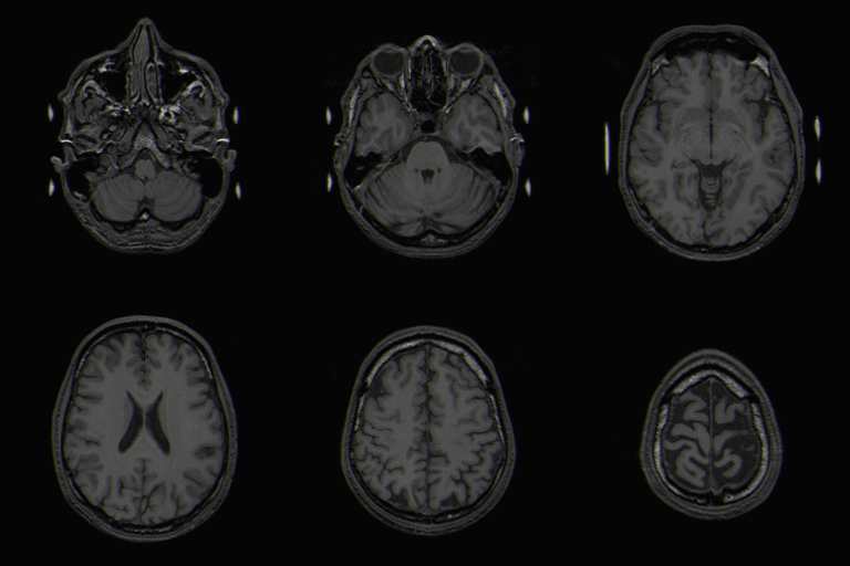
Facial Processing Patients with ASD show less interest in human faces. Normally people perform much better at tasks where they have to identify or match faces that are upright versus inverted. ASD children on the other hand, show a noticeably less pronounced 'inversion effect' as compared to the normal population. Some scientists speculate that this may be because children with ASD process faces similarly to regular objects rather than assigning particular significance to faces, thereby reducing the inversion effect. Another hypothesis, such as in Weak Coherence Theory is that ASD children may engage more in component processing rather than configural processing, thereby making the activation difference between processing upright faces versus inverted faces smaller. Differences in activation areas are in the frontal cortex and the amygdala, perhaps reflecting a differnce in processing the meaning and significance of faces. 2 Typically developing children also seem to activate the right prefrontal areas of the brain when doing self and other face processing while ASD children only activate this area when processing their own face.3A study examining the EEG differences in typical developing individuals and those with ASD during measures of face memory, cognitive functioning, and symptom levels found no significant differences. There were slight differences in speed on average. Authors hyopthesized that compensatory responses may have been used, and that ASD individuals with better language skills have a tendency to use those skills to scaffold face learning.12
One study found an association between amygdala response to emotional faces and social anxiety in those with ASD, and suggests that the level of social anxiety may mediate the neural response to emotional face perception. Greater social anxiety was associated with increased activation in the right amygdala and left middle temporal gyrus, and decreased activation in the fusiform face area. 13
Citations
