Category:Neuroimaging
Neuroimaging
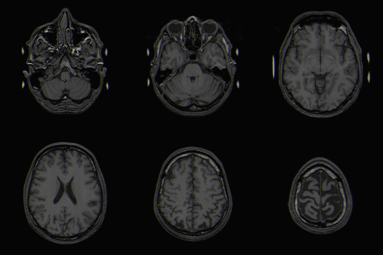
There are have been many differences observed between typically developing children and those with ASD in neuroimaging studies using fMRI, EEG, transcranial magnetic stimulation, EMG, and structural MRI. It has been hypothesized in the Mirror Neuron System Theory of Autism that the social/communication deficits in ASD arise because of differences in activation of the Mirror Neuron System (MNS), since the mirror neuron system plays an integral role in mediating understanding of emotional states of others. However, many different areas of the brain can participate in complex social/communicative tasks, making it difficult to single out just one neural area responsible for the widespread abnormalities in socialization and communication experienced by patients with ASD. Much of current neuroimaging research in ASD operates under one of two different camps that attribute autism to different areas of less activation in the brain.
- Theory of Mind-Patients with ASD show lower or no activation in midline structures as compared to a control groups in many tasks, lending credence to the 'Theory Of Mind' explanation for social/communication deficits in patients with ASD. In this theory, patients with ASD have lower activation of mirror neurons in the midline structures which are responsible for "reflecting" about others' emotions or wants1,. Thus, patients have social/communication deficits because they have are unable to process the meaning of other peoples' emotions, which suggests a problem in executive processing.
- Embodied Simulation Theory- Scientists who want to test this theory focus their research on shared circuits that are involved in both one's own emotions and other's emotions. Numerous studies have shown that when subjects witness a goal oriented task many different types of neurons are activated in addition to those in the midline structures. For example, when a person watches another person drink a glass of milk with an expression of disgust on his face, the premotor and parietal areas are activated for the action itself, the insula areas are activated in response to the emotion of disgust, and the secondary somatosensory area is activated for the sensations involved in the task. These are the same areas that would have been activated if we drank the milk and were ourselves disgusted. Thus, we are able to understand others' emotional states and intentions because contextual cues activate the same neural circuits that are activated when we perform the same task. However, patients with ASD show diminished activation of these areas as compared to control groups. So, in this theory, the social and communication difficulties arise in patients with ASD because these areas are less activated. Consequently, they have difficulty "understanding" another's actions.1
These two theories are not entirely incompatible and there have been some hypothesis on how the two theories could be integrated to provide a better explanation for the differences seen.
Neuroimaging research has shown that there are differences in both White Matter and Gray Matter tracts in the brain between typically developing children and those with ASD. Brain Disconnectivity in the above tracts is commonly seen.
Analytical Techniques
Pages in category "Neuroimaging"
The following 9 pages are in this category, out of 9 total.
